 payment page
DNA Replication is Semi-Conservative
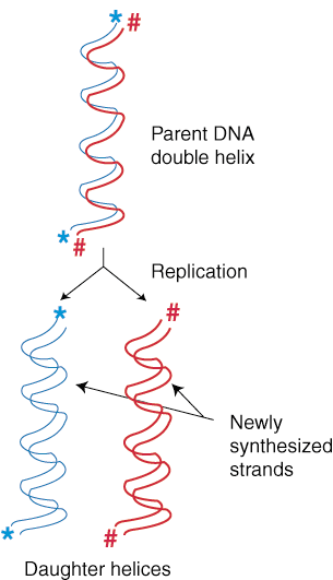
DNA replication of one helix of DNA results in two identical helices. If the original DNA helix is called the "parental" DNA, the two resulting helices can be called "daughter" helices. Each of these two daughter helices is a nearly exact copy of the parental helix (it is not 100% the same due to mutations). DNA creates "daughters" by using the parental strands of DNA as a template or guide. Each newly synthesized strand of DNA (daughter strand) is made by the addition of a nucleotide that is complementary to the parent strand of DNA. In this way, DNA replication is semi-conservative, meaning that one parent strand is always passed on to the daughter helix of DNA.
Replication Forks and Origins of Replication
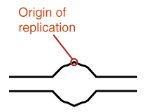
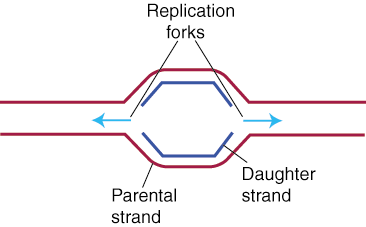
The first step in DNA replication is the separation of the two DNA strands that make up the helix that is to be copied. DNA Helicase untwists the helix at locations called replication origins. The replication origin forms a Y shape, and is called a replication fork. The replication fork moves down the DNA strand, usually from an internal location to the strand's end. The result is that every replication fork has a twin replication fork, moving in the opposite direction from that same internal location to the strand's opposite end. Single-stranded binding proteins (SSB) work with helicase to keep the parental DNA helix unwound. It works by coating the unwound strands with rigid subunits of SSB that keep the strands from snapping back together in a helix. The SSB subunits coat the single-strands of DNA in a way as not to cover the bases, allowing the DNA to remain available for base-pairing with the newly synthesized daughter strands.
As you can see in , when the two parent strands of DNA are separated
to begin replication, one strand is oriented in the 5' to 3' direction while the other
strand is oriented in the 3' to 5' direction. DNA replication, however, is inflexible:
the enzyme that carries out the replication, DNA polymerase, only functions in the 5'
to 3' direction. This characteristic of DNA polymerase means that the daughter strands
synthesize through different methods, one adding nucleotides one by one in the direction
of the replication fork, the other able to add nucleotides only in chunks. The first
strand, which replicates nucleotides one by one is called the leading strand; the
other strand, which replicates in chunks, is called the lagging strand.
The Leading and Lagging Strands
The Leading Strand
Since DNA replication moves along the parent strand in the 5' to 3' direction, replication can occur very easily on the leading strand. As seen in , the nucleotides are added in the 5' to 3' direction. Triggered by RNA primase, which adds the first nucleotide to the nascent chain, the DNA polymerase simply sits near the replication fork, moving as the fork does, adding nucleotides one after the other, preserving the proper anti-parallel orientation. This sort of replication, since it involves one nucleotide being placed right after another in a series, is called continuous.
The Lagging Strand
Whereas the DNA polymerase on the leading strand can simply follow the replication fork,
because DNA polymerase must move in the 5' to 3' direction, on the lagging strand the enzyme
must move away from the fork. But if the enzyme moves away from the fork, and the fork
is uncovering new DNA that needs to be replicated, then how can the lagging strand be
replicated at all? The problem posed by this question is answered through an ingenious
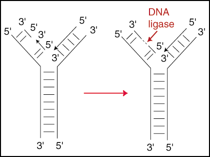
method. The lagging strand replicates in small segments, called Okazaki fragments.
These fragments are stretches of 100 to 200 nucleotides in humans (1000 to 2000 in bacteria)
that are synthesized in the 5' to 3' direction away from the replication fork. Yet while
each individual segment is replicated away from the replication fork, each subsequent
Okazaki fragment is replicated more closely to the receding replication fork than the
fragment before. These fragments are then stitched together by DNA ligase, creating
a continuous strand. This type of replication is called discontinuous
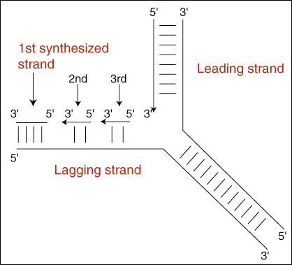
In figure above, we can also see how replication on the lagging strand remains slightly behind that on the leading strand. Because synthesis on the lagging strand takes place in a "backstitching" mechanism, its replication is slightly delayed in relation to synthesis on the leading strand. The lagging strand must wait for a patch of the parent helix to open up a short distance in front of the newly synthesized strand before it can begin its synthesis back to the end of the daughter strand. This "lag" time does not occur in the leading strand because it synthesizes the new strand by following right behind as the helix unwinds at the replication fork.