Did you know you can highlight text to take a note?
x
Suggestions
Use up and down arrows to review and enter to select.Please wait while we process your payment
If you don't see it, please check your spam folder. Sometimes it can end up there.
If you don't see it, please check your spam folder. Sometimes it can end up there.
Please wait while we process your payment
By signing up you agree to our terms and privacy policy.
Don’t have an account? Subscribe now
Create Your Account
Sign up for your FREE 7-day trial
Already have an account? Log in
Your Email
Choose Your Plan
Individual
Group Discount
Save over 50% with a SparkNotes PLUS Annual Plan!
 payment page
Purchasing SparkNotes PLUS for a group?
Get Annual Plans at a discount when you buy 2 or more!
Price
$24.99 $18.74 /subscription + tax
Subtotal $37.48 + tax
Save 25% on 2-49 accounts
Save 30% on 50-99 accounts
Want 100 or more? Contact us for a customized plan.
payment page
Your Plan
Payment Details
Payment Summary
SparkNotes Plus
You'll be billed after your free trial ends.
7-Day Free Trial
Not Applicable
Renews April 26, 2024 April 19, 2024
Discounts (applied to next billing)
DUE NOW
US $0.00
SNPLUSROCKS20 | 20% Discount
This is not a valid promo code.
Discount Code (one code per order)
SparkNotes PLUS Annual Plan - Group Discount
Qty: 00
SparkNotes Plus subscription is $4.99/month or $24.99/year as selected above. The free trial period is the first 7 days of your subscription. TO CANCEL YOUR SUBSCRIPTION AND AVOID BEING CHARGED, YOU MUST CANCEL BEFORE THE END OF THE FREE TRIAL PERIOD. You may cancel your subscription on your Subscription and Billing page or contact Customer Support at custserv@bn.com. Your subscription will continue automatically once the free trial period is over. Free trial is available to new customers only.
Choose Your Plan
For the next 7 days, you'll have access to awesome PLUS stuff like AP English test prep, No Fear Shakespeare translations and audio, a note-taking tool, personalized dashboard, & much more!
You’ve successfully purchased a group discount. Your group members can use the joining link below to redeem their group membership. You'll also receive an email with the link.
Members will be prompted to log in or create an account to redeem their group membership.
Thanks for creating a SparkNotes account! Continue to start your free trial.
We're sorry, we could not create your account. SparkNotes PLUS is not available in your country. See what countries we’re in.
There was an error creating your account. Please check your payment details and try again.
Please wait while we process your payment
Your PLUS subscription has expired
Please wait while we process your payment
Please wait while we process your payment
Each compartment and accessory organ serves a specific function. At each stage, the food is transformed into a slightly different form that allows it to be passed along to the next compartment. The coordination of functions is done through the nervous and endocrine systems. Through the process of digestion, food is transformed from large complex particles into basic elements.
The process of digestion begins in the mouth. Within the mouth lie the teeth, tongue and jaws. Through a chewing motion, the food is mechanically broken down between the teeth and mixed with saliva, which aids in chemical digestion. Upon stimulation, saliva is produced in the salivary glands and brought into the mouth. It contains salivary amylase, an enzyme that digests starch. Once the digestion in the mouth is completed, the first phase of swallowing is initiated. This stage is voluntary and is characterized by contraction of the muscles of the floor of the mouth and tongue that propel the food bolus into the pharynx.
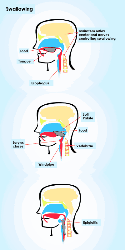
The role of the pharynx is to facilitate the passage of the food bolus into the esophagus. The pharynx is designed to direct the food bolus in this direction. It is here where the second phase of swallowing takes place. After the moistened food bolus is moved to the back of the mouth by the tongue, an involuntary swallowing reflex is triggered which prevents food from entering the respiratory tract. The tongue closes off the mouth, the soft palate blocks the nose, and the larynx rises such that the epiglottis closes off the trachea. Food then moves from the pharynx into the esophagus.
The esophagus is the first part of the digestive tract proper. It is also where the third phase of swallowing occurs. Approximately 10 inches in length, it consists of three tissue layers consistent with the rest of the gut. Once in the proximal portion of theesophagus, the muscles of peristalsis begin propelling the food bolus through the esophagus into the stomach.
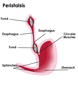
No further digestion takes place in this compartment. The pharynx and esophagus serve only as conduits for digestion.
The stomach is a C-shaped pouch that receives the food bolus from the esophagus. It aids both in mechanical and chemical digestion. Acting like a churn, the stomach mixes the food with gastric acid and breaks down the food into a milky substance known as chyme. The acid reduces the pH of the stomach, in the process allowing activation of an enzyme called pepsin. This starts the chemical digestive process.
Please wait while we process your payment