|
|
Amino Acids and Proteins
Functions of Proteins
Because proteins are a highly evolved and diverse class of molecules, they
perform endless tasks and functions within both plants and animals. They are
important in the biosyntheses of hormones, enzymes, and membrane channels and
pumps. In animals, proteins also function in the immune system and can be used
in the production of energy. In essence, proteins are the currency of life.
Biosyntheses: essential and nonessential amino acids (transanimation)
Since proteins constitute the majority of tissues in the body and since these
tissues are constantly in protein flux, proteins are degraded and synthesized
within all tissues on a regular basis. Some of the amino acids that are
degraded can be recycled by the liver and used again for other biosyntheses, but
a significant portion of this protein cannot be replaced.
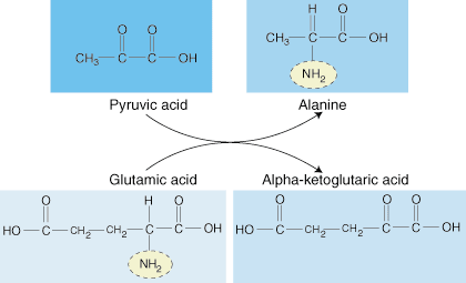
Through a process known as transamination, the liver synthesizes amino acids.
Figure 2.1: Transamination
During this reaction, an amino group from glutamic acid is transferred to an
alpha keto acid, which is a precursor for amino acid sythesis.
Aminotransferases, which are derived from vitamin B6, are the enzyme responsible
for the reaction. The amino acids that can be produced through transanimation
include alanine, arginine, asparagine, aspartic acid, cysteine, glutamic acid,
glutamine, glycine, proline, serine, and tyrosine. These are obviously the non-essential
amino acids, since they can be synthesized in the body.
Energy: ketogenic and glucogenic
When the body's energy sources are low, it begins to degrade proteins for use as
an alternative energy source. Amino acids can be classified as glucogenic or
ketogenic.
Glucogenic Amino Acids
Glucogenic amino acids can be degraded to pyruvate or an intermediate in the
Krebs Cycle. They are named glucogenic because they can produce glucose
under conditions of low glucose. This process is also known as
gluconeogenesis, or the production of "new glucose." Amino acids form
glucose through degradation to pyruvate or an intermediate in the Krebs Cycle.
Figure 2.2: Amino acid degradation to pyruvate
The intermediates can then be converted to oxaloacetate, the main
precursor for gluconeogenesis. The following amino acids are glucogenic:
alanine, cysteine, glycine, serine, threonine, tryptophan, asparagine,
aspartate, phenylalanine, tyrosine, isoleucine, methionine, threonine, valine,
arginine, glutamate, glutamine, histidine, and proline.
Ketogenic Amino Acids
In contrast, ketogenic amino acids can produce
ketones when energy sources are low. Some of these
amino acids are degraded directly to ketone bodies such as acetoacetate
(see ). They include leucine, lysine, phenylalanine,
tryptophan, and tyrosine. The other ketogenic amino acids can be converted to
acetyl CoA. Acetyl CoA has several different fates, one of which is the
conversion to acetoacetate. Although not a preferential energy source,
acetoacetate can be metabolized by the brain and muscle for energy when blood
glucose is low. Acetoacetate cannot be used in gluconeogenesis, since acetyl CoA
cannot be converted directly to oxaloacetate.
When energy sources are high, both glucogenic and ketogenic amino acids are
converted to fatty acids through the intermediate acetyl CoA. Other amino acids
that are degraded to intermediates in the Krebs Cycle are siphoned off into the
production of urea, a nitrogenous carboxyl compound that is filtered through the
kidneys and secreted in the urine.
Maintaining fluid balance
Blood proteins such as albumin and globulin function to maintain fluid balance
in the body. When the concentrations of proteins in the bloodstream are low,
the fluid in the blood (serum) begins to seep into surrounding tissue. Proteins
in the blood can counteract this effect by increasing the osmotic potential and
forcing fluid back into the bloodstream. Therefore, low amounts of protein in
the blood cause edema, a condition that is characterized by an abnormal amount
of fluid in the tissue and extracellular space. Edema is seen in starvation,
low calorie diets, and diseases like AIDS that decrease the amount of
circulating antibodies and albumin.
Hormones
Many hormones are composed of polypeptide chains. The beta cells in the
pancreas produce the peptide hormone insulin. Insulin facilitates glucose
uptake in cells and promotes the synthesis of glycogen and fatty acids.
Diabetics must inject the peptide hormone insulin because it will be degraded
into amino acids in the stomach and small intestine if taken orally. Other
examples of peptide signal molecules include neurotransmitters, a class of
molecules that are produced and released at nerve endings in the brain and
autonomic nervous system.
Enzymes
Enzymes are an entirely different class of proteins. Enzymes catalyze
biological reactions by increasing the reaction rates by factors of at least a
million. Since most reactions in the body proceed at imperceptible rates
without enzymes, it is critical that these proteins be present in sufficient
quantities for proper functioning of cells.
How are enzymes so efficient and fast? First, enzymes are highly specific for
their substrates. Substrates are any molecules for which the enzyme binds
preferentially or with a high affinity. Enzymes are able to bind specific
substrates because they form deep pockets or clefts that are complementary to
the three dimensional substrate conformations. Since the enzyme pocket is
complementary to the substrate, a large number of noncovalent, dipole-dipole,
and Van der Waal interactions can occur, favoring enzyme-substrate binding.
The second reason for the high catalytic rate of enzymes is that they are able
to stabilize transition state intermediates. By stabilizing these
intermediates, enzymes are able to decrease the activation energy required
for the reaction to occur. Upon reaching its high-energy transitional state,
the bound substrate can be easily converted to the cell's desired product, which
is then released to meet the needs of the cell. These reactions can take place
on the order of microseconds to nanoseconds. In fact, many enzymes are so
efficient and fast that they approach the diffusion-controlled limit, the rate
at which substrate diffusion cannot keep up with the rate at which the enzyme
catalyzes the reaction. Enzymes like these have reached catalytic perfection.
Besides their active sites, many enzymes have other locations or crevices where
molecules can bind. Regulatory sites, also called allosteric sites, are
places other than the active site of the enzyme that serve to regulate enzymatic
activity.
Allosteric sites as inhibitors
The end product in a series of reactions catalyzed by different enzymes at each
step may bind to the allosteric site and inhibit the activity of the first
enzyme in the pathway. When an inhibitory molecule binds to an allosteric
region on the enzyme it can cause the active site of the enzyme to close up and
become inactive.
Figure 2.3: Enzymatic negative feedback loop
This type of negative feedback is used to control levels of products from
reaching levels that are too high or unnecessary.
Allosteric sites as stimulators
Allosteric sites can also be areas that cause the stimulation of enzymatic
rates. When these allosteric sites are occupied, the enzyme's active site may
change in shape, becoming more efficient or receptive as a catalyst.
Covalent Modification
Further regulation of enzymes comes in the form of covalent modification.
Many enzymes are regulated by reversible attachment of phosphoryl groups to
serine andthreonine amino acid residues. Specific types of enzymes called
kinases phosphorylate add phosphoryl groups to other enzymes while phosphatases
remove phosphate groups. By adding just one covalent bond to the enzyme, its
activity can be drastically changed. For example, during levels of low blood
glucose, glucagon and epinephrine are secreted into the blood and bind to muscle
and brain cell receptors. Upon binding, these hormones cause a cascade of
effects within the cell, which results in the phosphorylation of a number of
proteins, including a series of enzymes involved in metabolism. All
phosphorylations act to increase the rate of enzymes involved in glycogen and
triglyceride degradation while inhibiting the rate of enzymes involved in
glycolysis and the Citric Acid Cycle. In effect, the addition of phosphate
groups to these enzymes causes the level of blood glucose to increase.
Membrane channels and pumps
Proteins are also abundant within biological membranes. Many cellular
receptors, channels, and pumps are bound to membranes. Since these proteins
span across a nonpolar environment, many of their residues facing this
environment are also nonpolar, allowing more favorable interactions to occur.
Both channels and pumps are involved in the regulation of fluids and ions within
and outside the cell. However, they differ in many key respects. Channels
allow ions to flow from an area of high concentration to an area of low
concentration. This is a completely passive process. Pumps, on the other hand,
force ions up their concentration gradient from a region of lower concentration
to a region of higher concentration. This process is called active
transport and usually requires the energy of adenosine triphosphate (ATP) in
order to overcome the energy barrier.
A classic example of a pump is the sodium potassium pump. Since during the
excitation of neurons sodium ions are constantly traversing into the cell and
potassium ions are leaving the cell, the resting levels of these ions must be
constantly restored. Unlike potassium, which is present in larger proportions
outside the cell, sodium is in much higher concentrations outside the cell. The
sodium potassium pump pushes sodium and potassium against their concentration
gradients by binding ATP and hyrolyzing it for energy.
Immune function
Proteins are also important in the immune response. There are two types of
important proteins the immune system uses to scan the vast network of molecules
in our body and determine self from nonself. In the first stage of the immune
response, the body recognizes circulating foreign particles (antigens) through
antibodies, which are produced by plasma cells (B lymphocytes). The ability of
plasma cells to produce millions of different antibodies is inherent from birth;
virtually any circulating antigen will be bound by its complementary antibody.
Once bound by antibody, the antigen can be either consumed by macrophages or be
further bound by mature plasma cells to stimulate the production of even more
antibodies. Mature cells are stimulated to divide and form clones, which act as
an immunological memory against further attack from identical antigens. Like
enzymes, the amino acid sequence of the antibody determines its specificity.
The whole recognition and response of plasma cells to a new infection is called
the humoral response.
The second stage of immune response is called the cellular immune response.
In the cellular response, T lymphocytes (killer T cells) bind to foreign
particles displayed on the surface of cells and destroy the contaminated cell.
Helper T cells also bind to the foreign particles displayed on the surface of
cells and stimulate the humoral response by helping plasma cells to proliferate.
Why is the cellular response necessary if antibodies can recognize antigens and
mark them for destruction? The answer lies in the fact that many of the viruses
and bacteria that invade the body are found in greater concentrations within
cells, preventing antibodies from reaching them. The immune system has adapted
to this problem by cutting some of the foreign particles up into peptides to be
displayed at the surface of the infected cell by a protein known as the major
histocompatibility complex (MHC). Killer and helper T cells are specialized for
recognizing peptides bound to these proteins, increasing the speed and
effectiveness of the immune response.
  Help |
Feedback |
Make a request |
Report an error |
Send to a friend
|
|