As we discussed in the cell cycle, before cells are allowed to enter the M phase they must meet certain cellular requirements. Entry into M phase is allowed by the formation of the mitotic cyclin-Cdk complex known as M phase-promoting factor that occurs as a cell cycle regulatory mechanism in the G2 phase.
The first phase of mitosis is called prophase. It follows G2, the final phase of interphase. A cell entering mitosis manifests a number of physical signs. Among these are condensation, or thickening, of chromosomes. Chromosome condensation is visible through a microscope and is required for subsequent chromosome separation during later stages of mitosis. Another physical characteristic of cells beginning mitosis is the sprouting of microtubules. Microtubules are protein filaments on which chromosomes migrate during mitosis.
Prophase
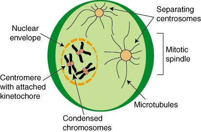
Figure 4.06: Prophase
As we discussed, prophase is marked by very thick and dense sister chromatids. At this phase, the sister chromatids are still enclosed in the cell nucleus within the nuclear envelope. The sister chromatids also contain a centromere, which is necessary in later phases for attachment to microtubules for migration. The centromere is a constricted the region that joins the sister chromatids together – it is the center of the X shape. Late in prophase, kinetochores assemble on the centromeres. Specialized microtubules, called kinetochore microtubules later attach to these sites. The network of cytoskeletal components begins to break down and the mitotic spindle forms. The mitotic spindle is an arrangement of microtubules that is responsible for aligning duplicated chromosomes in later phases.
As the nuclear envelope begins to break down into small vesicles, kinetochores become fully matured on the centromeres of the chromosomes. The disruption of the nuclear envelope allows for the mitotic spindles to gain access to the mature kinetochores. As the microtubules of the mitotic spindle enter the nuclear region, some attach to the kinetochores making them kinetochore microtubules. The remaining microtubules are called non-kinetochore microtubules. Sister chromatids are captured by microtubules. Once they have captured the sister chromatids, the kinetochore microtubules begin to exert force on the sister chromatids, moving them.
The next two major events that take place in mitosis are the alignment of sister chromatids at the center of the cell and the subsequent separation of sister chromatids to opposite mitotic spindle poles. These two events occur in metaphase and anaphase, respectively.
Metaphase
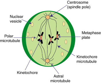
Figure 4.07: Metaphase
At the end of prophase, sister chromatids are being moved toward the center of the cell. Metaphase is marked by the alignment of the sister chromatids at the center of the cell, half way between each of the mitotic spindle poles. Movement is mediated by the kinetochore microtubules, which push and pull on the sister chromatids to align them into what is called the metaphase plate. Sister chromatids on the metaphase plate are held there tightly by pushing and pulling forces from the microtubules. Microtubules are dynamic molecules which allows them to hold the sister chromatids in place. The subunit of microtubules is a protein called tubulin and it is constantly added and removed from the ends of microtubules leading to a state of treadmilling.
Metaphase can occupy a large portion of the total time of mitosis because chromosome alignment at the center of the cell on the metaphase plate acts as a checkpoint for progression into the next phase, anaphase. Cells can arrest in metaphase for days until the sister chromatids are properly aligned and the cell enters anaphase.
Anaphase
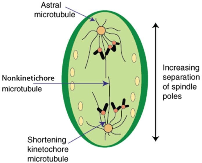
Figure 4.08: Anaphase
Entrance into anaphase is triggered by the inactivation of M phase-promoting factor that follows mitotic cyclin degradation. During anaphase, the kinetochore microtubules retract, increasing the separation of the chromosomes as they are moved further toward the opposite spindle poles. The extent of the separation of the poles varies from species to species. The entire duration of anaphase is relatively short, usually only lasting a few minutes.
The final two events of M phase are the re-forming of the nuclear envelope around the separated sister chromatids and the cleavage of the cell. These events occur in telophase and cytokinesis, respectively. In this section we will review the events that comprise these final phases of M phase.
Telophase
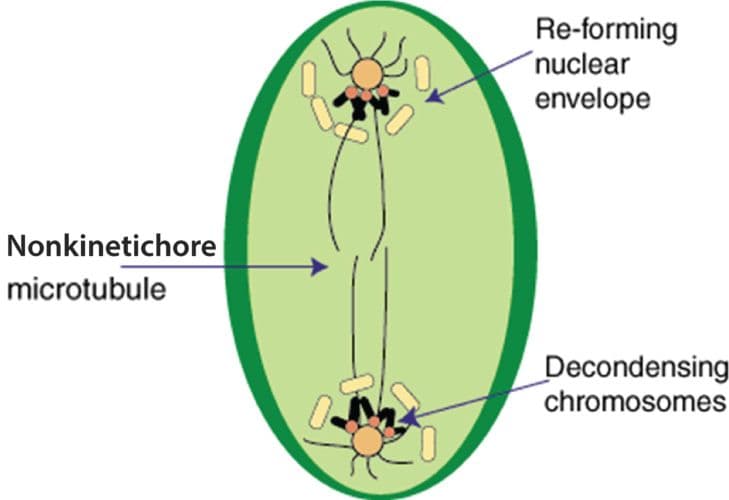
Figure 4.09: Telophase
Telophase is the final stage of mitosis. Its name derives from the Latin word telos which means end. During this phase, the chromosomes reach opposite poles. The small nuclear vesicles in the cell begin to re-form around the group of chromosomes at each end. As the nuclear envelope re-forms by associating with the chromosomes, two nuclei are created. Telophase is also marked by the dissolution of the kinetochore microtubules and the continued elongation of the nonkinetochore microtubules. As the nuclear envelopes re-form, the chromosomes begin to decondense and become more diffuse.
Cytokinesis
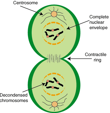
Figure 4.10: Cytokinesis
Cytokinesis is the process in which the cell actually divides into two. With the two nuclei already at opposite poles of the cell, the cell cytoplasm separates, and the cell pinches in the middle, ultimately leading to cleavage. In most cells, the mitotic spindle determines the site where the cell will begin to invaginate and split. The first signs of this puckering are usually visible sometime during anaphase.
Earlier we mentioned that in prophase, the cell's cytoskeleton becomes disassembled. The disassembled cytoskeletal filaments are used in a different way during cytokinesis. Cleavage occurs by the contraction of a thin ring of actin filaments that form the contractile ring. The contractile ring defines the cleavage line for the cell and forms a cleavage furrow. If the ring is not positioned at the center of the cell, an asymmetrical division takes place. The ring contracts and eventually pinches the cell until it separates into two independent daughter cells. In organisms with cell walls, the cytokinesis process is slightly different because the cytoplasm splits with the formation of a new cell wall.