


We will now begin our discussion of intracellular organelles. As we have mentioned, only eukaryotic cells have intracellular sub-divisions, so our discussion will exclude prokaryotic cells. We will also focus on animal cells, since plant cells have a number of further specialized structures. In this section we will discuss the importance of the cell nucleus, mitochondria, peroxisomes, endoplasmic reticulum, Golgi apparatus, and lysosome.
The Cell Nucleus
The cell nucleus is one of the largest organelles found in cells and also plays an important biological role. It composes about 10% of the total volume of the cell and is found near the center of eukaryotic cells. Its importance lies in its function as a storage site for DNA, our genetic material. The cell nucleus is composed of two membranes that form a porous nuclear envelope, which allows only select molecules in and out of the cell.
The DNA that is found in the cell nucleus is packaged into structures called chromosomes. Chromosomes contain DNA and proteins and carry all the genetic information of an organism. The nucleus is also the site of DNA replication and transcription.
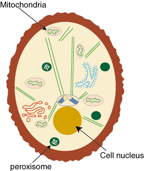
Figure 2.03: Location of the cell nucleus, mitochondria, and peroxisomes in a cell.
Mitochondria
Mitochondria, with their specialized double-membrane structure, generate adenosine triphosphate (ATP), a molecule that provides organisms with energy.
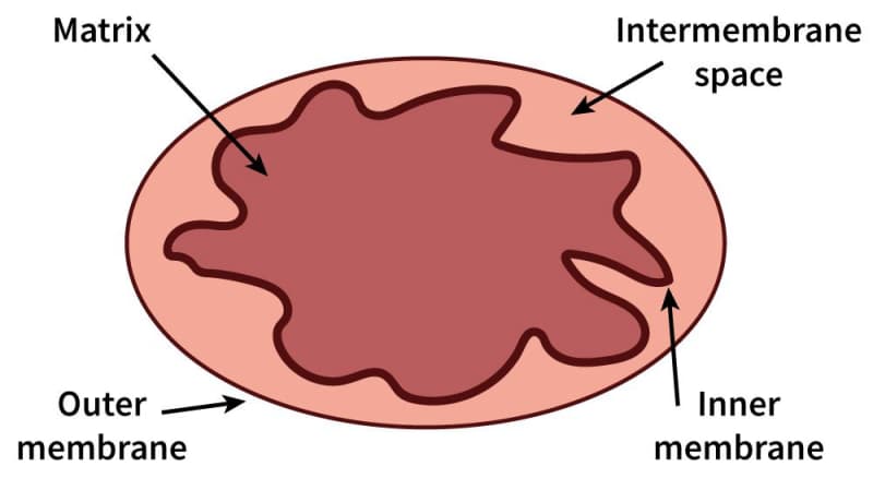
Figure 2.04: Mitochondrial structure
The outer and inner membranes of mitochondria form two sub-compartments: the internal matrix space and the intermembrane space. Mitochondria synthesize ATP with energy supplied by the electron transport chain and a process called oxidative phosphorylation.
Peroxisomes
Peroxisomes are single-membrane structures found in all eukaryotic cells. They are small, membrane-bound structures that use molecular oxygen to oxidize organic molecules. The structure is one of the major oxygen-utilizing organelles, the other being mitochondria. Peroxisomes contain oxidative enzymes and other enzymes that help produce and degrade hydrogen peroxide. Because of their varying enzymatic compositions, peroxisomes are diverse structures. Their main function is to help break down fatty acids.
The Endoplasmic Reticulum
The endoplasmic reticulum, or ER, is a very important cellular structure because of its function in protein synthesis and lipid synthesis. For example, the ER is the site of production of all transmembrane proteins. Since nearly all proteins that are secreted from a cell pass through it, the ER is also important in cellular trafficking. In addition to these major roles, the ER plays a role in a number of other biological processes. There are two different types of ER: smooth ER and rough ER (RER).
The rough ER has its name because it is coated with ribosomes, the structures most directly responsible for carrying out protein synthesis. Ribosomes are composed of ribosomal RNA (rRNA) and proteins. Together, these molecules guide the bonding of amino acids together to form polypeptide chains. Smooth ER lacks these ribosomes and is more abundant in cells that are specific for lipid synthesis and metabolism.
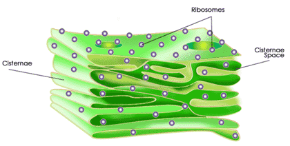
Figure 2.05: The Endoplasmic Reticulum
In addition to protein and lipid synthesis, the ER also conducts post-synthesis modifications. One such modification involves the addition of carbohydrate chains to the proteins, which assist in cell communication. Another major modification is called protein folding, whose name is rather self-explanatory. Another role of the ER is to capture calcium for the cell from the cytosol. Finally, the ER can secrete proteins into the cell that are usually destined for the Golgi apparatus.
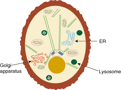
Figure 2.06: The location of the Endoplasmic Reticulum, Golgi apparatus, and lysosome in a eukaryotic cell.
The Golgi Apparatus
The Golgi apparatus is usually located near the cell nucleus. It is composed of a series of layers called Golgi stacks. Proteins from the ER always enter and exit the Golgi apparatus from the same location. The cis face of the Golgi is where proteins enter. A protein will make its way through the Golgi stacks to the other end called the trans face where it is secreted to other parts of the cell.
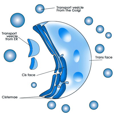
Figure 2.07: Structure of the Golgi Apparatus
In the Golgi apparatus, more carbohydrate chains are added to the protein while other chains are removed. This facilitates the folding and joining of polypeptides to form the tertiary and quaternary structure of a protein. The Golgi stacks also sort proteins for secretion. After sorting, the membrane of the Golgi buds off, forming secretory vesicles that transport proteins to their specific destination in the cell. A protein's destination is often signaled with a specific amino acid sequence at its end. A protein secretion most often travels back to the ER, to the plasma membrane where it can become a transmembrane protein, or to the next structure we will discuss, the lysosomes.
Lysosomes
Lysosomes are sites of molecular degradation found in all eukaryotic cells. They are small, single-membrane packages of acidic and hydrolytic enzymes that digest molecules and are found throughout eukaryotic cells. As such, lysosomes are a sort of cellular "garbage can," getting rid of cellular debris. Proteins that are not correctly folded or have significant mutations can be secreted to the lysosomes and be degraded instead of taking up space in the cell.
Molecules from outside a cell can be taken in through a process called endocytosis. In this process, the cell membrane invaginates, forming a vesicle containing the transported molecule that will eventually reach a lysosome. The reverse of endocytosis is exocytosis. In this process, molecules within a cell are secreted into lysosome. After reaching the lysosomes, the molecules are secreted from a cell in membrane vesicles.