The brain is an essential part of the nervous system, a complex, highly coordinated network of tissues that communicate through electrochemical signals. We use our brains in virtually everything we do, from keeping our heart beating to deducing the existence of black holes. Within our brains lie our deepest secrets, our earliest memories, our most amazing capabilities, and the keys to the mystery of consciousness itself. The brain is divided into three major regions: the hindbrain, the midbrain, and the forebrain.
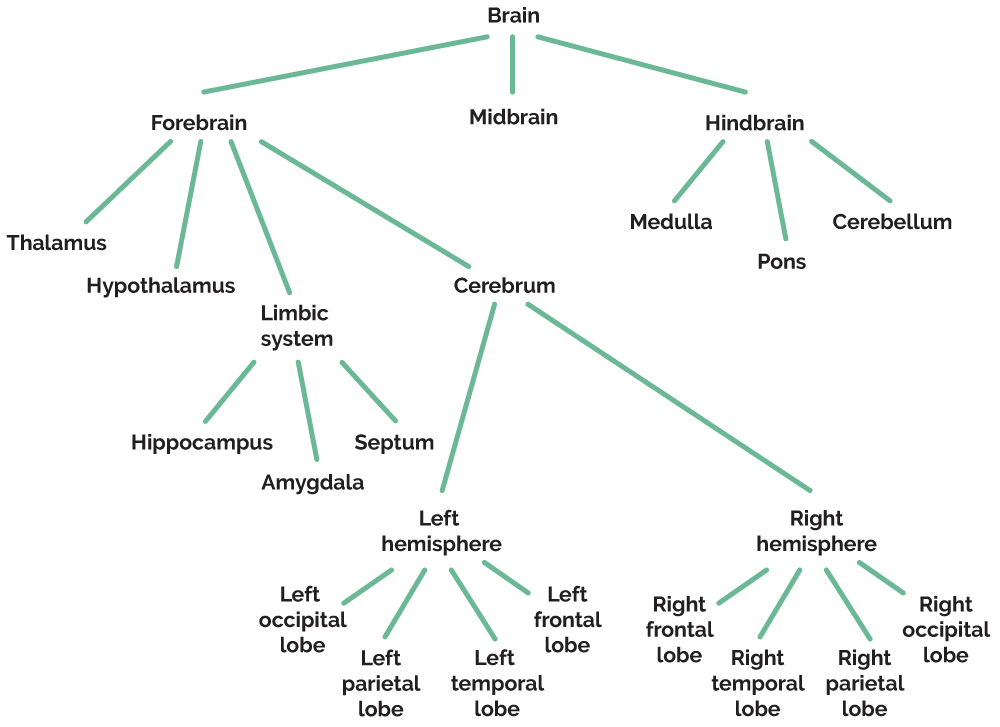
The Hindbrain
The hindbrain, at the back of the brain, is responsible for regulating many of the body’s most vital functions, such as breathing, heart rate, and balance. The hindbrain includes the medulla oblongata, the pons, and the cerebellum. An important part of the hindbrain is the brainstem, which connects the brain to the spinal cord and transmits information between the brain and the rest of the body. The brainstem is composed of the medulla oblongata, pons (both part of the hindbrain), and the midbrain (which belongs to a separate brain region).
The medulla oblongata, located at the lowest part of the brainstem, controls autonomic functions—those outside of conscious control—such as heart rate, breathing, and reflexes like sneezing and swallowing. In essence, the medulla oversees the body’s most basic survival functions.
The pons, which sits right under the midbrain and right above the medulla, serves as a “bridge” that connects the lower part of the brainstem to the cerebral cortex. The pons regulates sleep-wake cycles and manages pain signals. It also works with other brain structures to regulate hearing, balance, and face and eye movements.
The cerebellum, located behind the brainstem, is essential for coordinating balance, posture, and voluntary movements. While traditionally associated with motor control, research shows that the cerebellum also plays a role in regulating emotion and decision-making. It is vital for procedural memories—the memories that help us perform skills such as riding a bike or playing the piano. Damage to the cerebellum can impair fine motor skills, making tasks like playing an instrument or typing more difficult.
The brainstem also contains the reticular activating system (RAS), which is a network of neurons extending through the brainstem and midbrain with connections to the cerebral cortex. The RAS regulates arousal, consciousness, attention, and motivation. It helps people adapt to ongoing background noise and selectively notice important changes in the environment. It is the system that allows a parent to wake to the sound of their crying baby when they may otherwise sleep through other sounds, or to no longer notice the noise of a running dishwasher or air conditioner. Damage to the RAS is associated with comas, as it is crucial for maintaining wakefulness and consciousness.
The Midbrain
The midbrain, or mesencephalon, is an important part of the brainstem, located between the hindbrain and forebrain. It plays a key role in integrating sensory information and coordinating motor movements while also contributing to alertness and arousal.
One of the midbrain’s primary roles is processing sensory input, particularly visual and auditory stimuli. Structures in the midbrain control reflexive movements, such as turning your head toward sudden noise or following a moving object with your eyes. The midbrain ensures that sensory information is rapidly processed, allowing quick responses to environmental changes. Additionally, the midbrain regulates motor control by working closely with the basal ganglia and cerebellum. This coordination allows for smooth and deliberate movement, demonstrating how the midbrain integrates sensory and motor systems.
The midbrain also contains part of the reticular formation described above, the network of neurons involved in maintaining alertness, wakefulness, and consciousness. By managing attention and arousal, the midbrain supports higher cognitive functions, such as problem-solving and decision-making.
The Limbic System
The limbic system is a group of interconnected structures in the brain that play a central role in emotion, motivation, and memory. It serves as a bridge between the brainstem and the higher-order thinking areas of the cerebral cortex, helping humans respond to emotional stimuli, form memories, and regulate motivated behaviors like hunger, thirst, and reproduction. Some key structures of the limbic system include the thalamus, the hypothalamus, the amygdala, and the hippocampus.
The thalamus acts as a relay station for all sensory information except smell-related data, directing it to the appropriate areas of the cerebral cortex for processing. While not directly responsible for emotions, the thalamus plays a critical role in integrating sensory data with emotional responses, enabling individuals to react to their environment effectively. For example, when you hear a loud noise, the thalamus quickly relays that auditory information to the appropriate brain regions, allowing a rapid emotional or defensive response.
The hypothalamus regulates essential autonomic functions like hunger, thirst, body temperature, and circadian rhythms. It also controls the endocrine system through its connection to the pituitary gland, which is the “master gland” that regulates various hormonal fluctuations and controls the activity of other endocrine glands throughout the body. The hypothalamus controls the release of hormones from the pituitary gland, linking the nervous system to the endocrine system. The hypothalamus plays a key role in motivation, driving behaviors necessary for survival, such as eating, drinking, and reproduction. Additionally, it is involved in emotional responses, such as aggression and pleasure.
The amygdala is often referred to as the brain’s emotional center, as it plays a crucial role in processing fear, anger, and other strong emotions. It evaluates stimuli for emotional significance and initiates appropriate responses, such as fight-or-flight reactions to perceived threats. Beyond fear, the amygdala is also involved in forming and retrieving emotional memories, which helps individuals remember emotionally-charged experiences, such as joyful celebrations or traumatic events. Damage to the amygdala can impair the ability to recognize emotions in others or appropriately respond to fear-inducing situations.
The hippocampus is essential for memory formation, particularly in converting short-term memories into long-term memories. It does not store memories itself but acts as a hub that organizes and consolidates information for storage in other areas of the brain. The hippocampus is also involved in spatial navigation, helping individuals mentally map their surroundings. Damage to the hippocampus can lead to severe memory deficits, such as the inability to form new memories.
The Cerebral Cortex
The cerebral cortex is the outermost layer of the brain, responsible for higher-order functions such as thinking, perception, language, and voluntary movement. It is part of the cerebrum, the largest brain structure. The cerebral cortex is divided into four lobes, each with specialized functions. Each lobe contains a primary cortex that handles sensory or motor functions and several association areas that handle higher-level processing specific to the functions of that lobe. Association areas integrate and process information from different sensory and motor areas rather than directly controlling movement or receiving sensory input. These areas allow for complex cognitive tasks such as reasoning, problem-solving, language, memory, and decision-making.
The four lobes of the cerebral cortex are as follows:
The occipital lobe, located at the back of the brain, contains the primary visual cortex, which handles visual information. This lobe contains association areas involved in processing and interpreting visual information.
The parietal lobe, located near the back crown of the brain, contains the primary somatosensory cortex, which handles information related to the sense of touch. The parietal lobe also plays a part in sensing body position and integrating visual information. This lobe has association areas that integrate sensory information from the somatosensory cortex to help with spatial awareness and attention.
The temporal lobe, located on the sides of the brain underneath the parietal lobe, contains the primary auditory cortex, which is involved in processing auditory information. This lobe contains association areas related to language comprehension and memory, such as Wernicke’s area for understanding speech. Damage to Wernicke’s area can lead to Wernicke’s aphasia, a condition where individuals may speak fluently but struggle to comprehend language and produce meaningful sentences.
The frontal lobe, located at the front of the brain just behind the forehead, contains the primary motor cortex, which controls muscle movement. It is also responsible for many higher-level cognitive functions, such as planning, decision-making, and problem-solving. The frontal lobe includes association areas such as Broca’s area, which influences speech production as well as association areas responsible for higher-order thinking, decision-making, personality, and planning. Damage to Broca’s area can lead to Broca’s aphasia, where individuals can understand speech but struggle to produce it. Both Broca’s area and Wernicke’s area are primarily associated with the left hemisphere, as language processing is associated with the left hemisphere.
Brain Hemispheres
The cerebrum is the biggest part of the brain; it controls complex processes such as abstract thought, memory, and learning. The corpus callosum is a band of fibers that runs along the cerebrum from the front of the skull to the back, dividing the cerebrum into two halves or hemispheres. The left and right hemispheres of the brain are the two halves of the cerebrum, each responsible for different functions and processing styles. This division is known as hemispheric specialization.
The left hemisphere is typically associated with analytical and logical thinking, language processing, and mathematical skills, making it crucial for tasks such as reading, writing, and verbal communication. Remember that Broca’s area, responsible for speech production, and Wernicke’s area, responsible for language comprehension, are two critical language areas located in the left hemisphere. In addition to language, the left hemisphere is involved in mathematical calculations and logical problem-solving, making it crucial for tasks that require orderliness and precision.
The right hemisphere is associated with creativity, spatial awareness, and the interpretation of nonverbal cues, playing a vital role in artistic expression and emotional processing. It plays a key role in making inferences, modulating speech, processing perceptual tasks, and orchestrating self-awareness. The right hemisphere helps interpret implied meanings and context, enabling a nuanced understanding of language and situations. While not responsible for speech production, the right hemisphere contributes to the emotional tone of speech, helping convey meaning beyond words. It also processes visual and spatial information, such as recognizing faces, understanding maps, and navigating spaces. Finally, the right hemisphere is involved in recognizing one’s own body, emotions, and presence in space, contributing to a sense of self.
Although the hemispheres work together and communicate through the corpus callosum, individuals may exhibit a dominant hemisphere, influencing their cognitive strengths and preferences. Lateralization refers to the fact that the right and left hemispheres of the brain regulate different functions. The left hemisphere specializes in verbal processing tasks such as writing, reading, and talking. The right hemisphere specializes in nonverbal processing tasks such as playing music, drawing, and recognizing childhood friends. Roger Sperry, Michael Gazzaniga, and their colleagues conducted some of the early research in lateralization. They examined people who had undergone split-brain surgery, an operation to cut the corpus callosum and separate the two brain hemispheres. Doctors sometimes use split-brain surgery as a treatment for epileptic seizures. Their research revealed how the left and right hemispheres operate independently when they are no longer able to communicate with each other, providing insight into how lateralization affects perception and behavior.
One important concept related to brain function is contralateral hemispheric organization, which refers to how each hemisphere controls the opposite side of the body. Because of the organization of the nervous system, the left hemisphere of the brain controls the functioning of the right side of the body. Likewise, the right hemisphere controls the functioning of the left side of the body. Vision and hearing operate a bit differently. What the left eye and right eye see goes to the entire brain. However, images in the left visual field stimulate receptors on the right side of each eye, and information goes from those points to the right hemisphere. Similarly, information in the right visual field ends up in the left hemisphere. In the case of auditory information, both hemispheres receive input about what each ear hears. However, information first goes to the opposite hemisphere. If the left ear hears a sound, the right hemisphere registers the sound first. The fact that the brain’s hemispheres communicate with opposite sides of the body does not affect most people’s day-to-day functioning because the two hemispheres constantly share information via the corpus callosum. However, severing the corpus callosum and separating the hemispheres causes impaired perception.
Split-Brain Studies
If a researcher presented a picture of a Frisbee to a split-brain patient’s right visual field, information about the Frisbee would go to his left hemisphere. Because language functions reside in the left hemisphere, he’d be able to say that he saw a Frisbee and describe it. However, if the researcher presented the Frisbee to the patient’s left visual field, information about it would go to his right hemisphere. Because his right hemisphere can’t communicate with his left hemisphere when the corpus callosum is cut, the patient would not be able to name or describe the Frisbee.
The same phenomenon occurs if the Frisbee is hidden from sight and placed in the patient’s left hand, which communicates with the right hemisphere. When the Frisbee is in the patient’s left visual field or in his left hand, the patient may not be able to say what it is, although he would be able to point to a picture of what he saw. Picture recognition requires no verbal language and is also a visual-spatial task, which the right hemisphere controls.
Brain Plasticity
The brain's ability to reorganize itself by forming new neural connections is called brain plasticity, also known as neuroplasticity. There are two types of plasticity: structural plasticity, in which the brain’s physical structure changes in response to learning and experience, and functional plasticity, which refers to the brain’s ability to move functions from a damaged area to undamaged areas. Plasticity is most robust during early development periods, especially in childhood, when the brain is still learning new skills such as language or motor functions. Plasticity continues throughout life, just to a lesser degree.
Brain plasticity plays a crucial role in rehabilitation after brain injuries, such as strokes, where undamaged regions of the brain can compensate for lost functions like movement or speech. Research on brain plasticity uses tools like functional magnetic resonance imaging (fMRI) and electroencephalography (EEG) to observe how neural activity changes with experience or after damage. Case studies of individuals with brain injuries have provided valuable insight into how the brain adapts. Understanding brain plasticity is key in developmental psychology, where it explains how children learn new skills, and in cognitive psychology, as it relates to how the brain adapts and reorganizes through learning and memory formation.
Studying the Brain
To examine the brain’s functions, researchers have to study a working brain, which means they can’t use cadavers. Invasive studies, in which researchers put instruments into the brain, can’t be done in humans, though they can be done occasionally during medically-necessary brain surgery. Researchers usually use invasive techniques in animal studies. There are two main types of invasive animal studies—lesioning studies and electric stimulation:
Lesioning studies: Researchers use an electrode and an electric current to burn a specific, small area of the brain.
Electric stimulation of the brain: Researchers activate a particular brain structure by using a weak electric current sent along an implanted electrode.
Because they cannot use such invasive techniques on humans, researchers study human brains in two ways:
They examine people with brain injuries or diseases and see what they can and can’t do.
They use electroencephalographs (EEGs), which can record the overall electrical activity in the brain via electrodes placed on the scalp.
Recently, high-tech innovations have made studying human brains easier. Researchers use three types of imaging equipment to study the brain:
Computerized tomography (CT): In CT, a number of x-rays are taken of the brain from different angles. A computer then combines the x-rays to produce a picture of a horizontal slice through the brain.
Magnetic resonance imaging (MRI): Both brain structure and function can be visualized through MRI scans, which are computer-enhanced pictures produced by magnetic fields and radio waves.
Positron emission tomography (PET): For PET scans, researchers inject people with a harmless radioactive chemical, which collects in active brain areas. The researchers then look at the pattern of radioactivity in the brain, using a scanner and a computer, and figure out which parts of the brain activate during specific tasks, such as lifting an arm or feeling a particular emotion.