


Thanks to the nose, ears, eyes, tongue, and skin, we can imagine a day at the beach: glimmering blue sky, salty water, warm sand, and crying seagulls. Our knowledge of the world depends on the senses: smell, hearing, vision, taste, touch, position, movement, and balance. If someone bounces a basketball, our eyes and ears pick up stimuli such as light and sound waves and send neural signals to the brain. This process called “sensation” occurs when physical energy from objects in the world or in the body stimulates the sense organs.
The Senses
Sensation is the process by which physical energy from objects in the world or in the body stimulates the sense organs. The brain interprets and organizes this sensory information in a process called perception.
Measuring the Senses
Psychologists assess the acuity of the senses in three ways:
- Measuring the absolute threshold
- Measuring the difference threshold
- Applying signal detection theory
The absolute threshold is the minimum amount of stimulation required for a person to detect the stimulus 50 percent of the time. The difference threshold is the smallest difference in stimulation that can be detected 50 percent of the time. The difference threshold is sometimes called the just noticeable difference (JND), and it depends on the strength of the stimulus.
Example: If someone were comparing two weak stimuli, such as two very slightly-sweet liquids, he’d be able to detect quite a small difference in the amount of sweetness. However, if he were comparing two intense stimuli, such as two extremely sweet liquids, he could detect only a much bigger difference in the amount of sweetness.
Weber’s Law
Ernst Weber, a 19th-century psychologist, proposed a principle demonstrating the fact that we can’t detect the difference between two stimuli unless they differ by a certain proportion and that this proportion is constant. In other words, the just noticeable difference for a stimulus is in a fixed proportion to the magnitude of a stimulus. Weber’s law holds true except in the most extreme kinds of stimulation.
Researchers use signal detection theory to predict when a weak signal will be detected. This theory considers the fact that the ability to detect a signal depends not only on the strength of the signal but also on the perceiver’s experience, motivation, expectation, and degree of alertness. Different people respond differently to the same signal, and the same person may detect a particular signal at one time but not another. Furthermore, people can often detect one type of signal in a sensory modality such as hearing or vision but be oblivious to other types of signals in the same sensory modality.
Sensory Adaptation
When people walk into a restaurant, they probably notice food smells right away. However, as they sit in the restaurant, the smells gradually become less noticeable. This phenomenon occurs because of sensory adaptation. Sensory adaptation is the decrease in sensitivity to an unchanging stimulus. The smells don’t disappear—the people just become less sensitive to them.
Development of the Senses
Babies have all the basic sensory abilities and many perceptual skills, but these abilities develop and grow more sensitive over time. Babies can recognize the difference between a human voice and other sounds, and they can locate a sound’s origin. They can recognize the difference between smells and, very early on, can recognize their mother’s particular smell. As for taste, they can differentiate between sweet and salty. Babies also have fairly adept visual abilities. Soon after birth, they can distinguish objects of different colors and sizes. When they are just a few weeks old, they begin to differentiate among contrasts, shadows, and patterns, and they can perceive depth after just a few months.
Sensitive Periods
Even innate perceptual skills need the right environment to develop properly. A lack of certain experiences during sensitive periods of development will impair a person’s ability to perceive the world.
Example: People who were born blind but regain their vision in adulthood usually find the visual world confusing. Since these adults were blind in infancy, they missed the sensory experiences necessary for their visual system to develop fully.
Sensory Interaction
The sensory systems all work together in a process called sensory interaction, where different senses influence or enhance other senses. For example, the taste of food is influenced by how it smells, which is why food may seem bland or flavor-muted when someone is congested. Another example of sensory interaction involves vision and balance; vision interacts with the vestibular sense (balance), which explains why balancing becomes more difficult with one’s eyes closed. Sensory interaction can also be demonstrated through the McGurk effect, a phenomenon in which visual information, such as lip movement, can alter auditory perception. If, for instance, a person watches a video of someone saying “fat” but hears an audio track playing the word “bat,” the brain may combine the two inputs to perceive a third sound, for example, “dat.” This demonstrates how the brain integrates conflicting visual and auditory information, leading to a different perception of sound altogether.
Synesthesia
The experience of synesthesia provides abnormal but informative examples of sensory interaction. Synesthesia is a rare condition (occurring in about 1 in 2,000 to 1 in 20,000 people) in which stimulation of one sensory system automatically causes one to experience the activation of another sensory system. For example, someone with synesthesia might “see” colors when they listen to music, or they may associate certain sounds with specific flavors. There are multiple forms of synesthesia, but the most common types are grapheme-color synesthesia (where letters or numbers are perceived as specific colors) and sound-to-color synesthesia (where sounds trigger visual experiences of color). While the exact causes of synesthesia aren’t fully understood, it is believed to result from cross-activation between areas of the brain that are responsible for different sensory processes. This might involve increased connectivity between sensory regions. Synesthesia appears to run in families, which suggests a genetic component. However, the exact type can vary from person to person. Synesthesia can also be acquired through brain damage, sensory deprivation, or drugs. However, synesthesia that is acquired is less consistent than synesthesia that is present from birth.
Vision
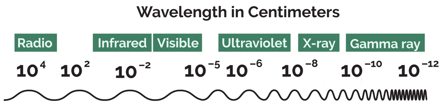
Researchers have studied vision more thoroughly than the other senses. Because people need sight to perform most daily activities, the sense of sight has evolved to be highly sophisticated. Vision, however, would not exist without the presence of light. Light is electromagnetic radiation that travels in the form of waves. Light is emitted from the sun, stars, fire, and lightbulbs. Most other objects just reflect light.
People experience light as having three features: color, brightness, and saturation. These three types of experiences come from three corresponding characteristics of light waves:
|
Light Feature |
Light Wave Characteristic |
|
Color |
Wavelength |
|
Brightness |
Amplitude |
|
Saturation |
Complexity |
The color or hue of light depends on its wavelength, the distance between the peaks of its waves.
The brightness of light is related to intensity or the amount of light an object emits or reflects. Brightness depends on light wave amplitude, the height of light waves. Brightness is also somewhat influenced by wavelength. Yellow light tends to look brighter than reds or blues.
Saturation or colorfulness depends on light complexity, the range of wavelengths in light. The color of a single wavelength is pure spectral color. Such lights are called fully saturated. Outside a laboratory, light is rarely pure or of a single wavelength. Light is usually a mixture of several different wavelengths. The greater number of spectral colors in a light, the lower the saturation. Light of mixed wavelengths looks duller or paler than pure light.
Rainbows and Lights
White light: Completely unsaturated. It is a mixture of all wavelengths of light.
The visible spectrum: Includes the colors of the rainbow, which are red, orange, yellow, green, blue, indigo, and violet.
Ultraviolet light: The kind of light that causes sunburns. It has a wavelength somewhat shorter than the violet light at the end of the visible spectrum.
Infrared radiation: Has a wavelength somewhat longer than the red light at the other end of the visible spectrum.
The process of vision cannot be understood without some knowledge about the structure of the eye:
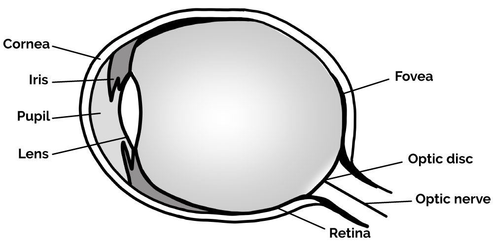
The cornea is the transparent, protective outer membrane of the eye.
The iris, the colored part of the eye, is a ring of muscle.
The iris surrounds an opening called the pupil, which can get bigger or smaller to allow different amounts of light through the lens to the back of the eye. In bright light, the pupil contracts to restrict light intake; in dim light, the pupil expands to increase light intake.
The lens, which lies behind the pupil and iris, can adjust its shape to focus light from objects that are near or far away. This process is called accommodation.
Light passing through the cornea, pupil, and lens falls onto the retina at the back of the eye. The retina is a thin layer of neural tissue. It contains photoreceptor cells (rods and cones) that detect light and color. The retina converts light into neural signals (a process called transduction), which are sent to the brain via the optic nerve. The image that falls on the retina is always upside down.
The center of the retina, the fovea, is where vision is sharpest. This explains why people look directly at an object they want to inspect. This causes the image to fall onto the fovea, where vision is clearest.
Accommodation and Focus
Visual stimuli are focused onto the retina by the lens through a process called accommodation, which involves the lens changing shape to focus light from objects at various distances. In accommodation, the ciliary muscles adjust the curvature of the lens, making it thicker to focus on nearby objects and thinner for distant objects. When the lens cannot properly focus light onto the retina, vision problems such as nearsightedness (myopia) or farsightedness (hyperopia) can occur. Nearsightedness happens when the lens focuses light in front of the retina, making distant objects appear blurry. In contrast, farsightedness occurs when the lens focuses light behind the retina, making nearby objects difficult to see clearly.
Rods and Cones
The retina has millions of photoreceptors called rods and cones. Photoreceptors are specialized cells that respond to light stimuli. There are many more rods than cones. The long, narrow cells, called rods, are highly sensitive to light and allow vision even in dim conditions. There are no rods in the fovea, which is why vision becomes hazy in dim light. However, the area just outside the fovea contains many rods, and these allow peripheral vision.
Because rods are so sensitive to light, in dim-lighting conditions, peripheral vision is sharper than direct vision.
Example: People can often see a star in the night sky if they look a little to the side of the star instead of directly at it. Looking to the side utilizes peripheral vision and makes the image of the star fall onto the periphery of the retina, which contains most of the rods.
Cones are cone-shaped cells that can distinguish between different wavelengths of light, allowing people to see in color. Cones don’t work well in dim light, however, which is why people have trouble distinguishing colors at night. The fovea has only cones, but as the distance from the fovea increases, the number of cones decreases.
|
Feature |
Rods |
Cones |
|
Shape |
Long and narrow |
Cone-shaped |
|
Sensitivity to light |
High: help people to see in dim light |
Low: help people to see in bright light |
|
Help Color Vision |
No |
Yes |
|
Present in Fovea |
No |
Yes |
|
Abundant in Periphery of Retina |
Yes |
No |
|
Allow Peripheral Vision |
Yes |
No |
Adaptation to Light
Dark adaptation is the process by which receptor cells sensitize to light, allowing clearer vision in dim light. Light adaptation is the process by which receptor cells desensitize to light, allowing clearer vision in bright light.
Connection to the Optic Nerve
Rods and cones connect through synapses to bipolar cells, which act as intermediaries by transmitting visual information from the rods and cones to ganglion cells, which are the final output neurons of the retina. The axons of all the ganglion cells in the retina come together to make up the optic nerve. The optic nerve connects to the eye at a spot in the retina called the optic disk. The optic disk is also called the blind spot because it has no rods or cones. Any image that falls on the blind spot disappears from view, since no photoreceptor cells are present to capture visual information. We do not usually notice the visual gap because the brain compensates by filling in the missing information, allowing us to perceive a continuous, complete image of the world.
Transmission of Visual Information
Visual information travels from the eye to the brain as follows:
- Light reflected from an object hits the retina’s rods and cones.
- Rods and cones send neural signals to the bipolar cells.
- Bipolar cells send signals to the ganglion cells.
- Ganglion cells send signals through the optic nerve to the brain.
Bipolar and ganglion cells gather and compress information from a large number of rods and cones. The rods and cones that send information to a particular bipolar or ganglion cell make up that cell’s receptive field.
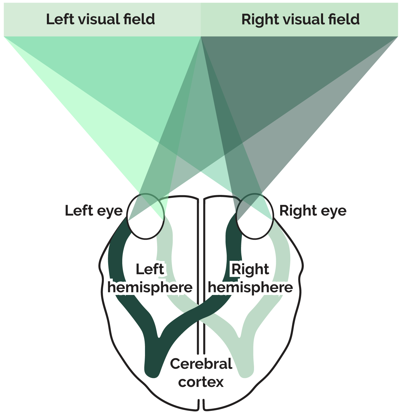
Ganglion cell axons from the inner half of each eye cross over to the opposite half of the brain. This means that each half of the brain receives signals from both eyes. Signals from the left side of the eyes go to the left side of the brain, and signals from the right sides of the eyes go to the right side of the brain. The diagram below illustrates this process.
Visual Processing in the Brain
After being processed in the thalamus and different areas of the brain, visual signals eventually reach the primary visual cortex in the occipital lobe of the brain’s cerebrum. In the 1960s, David Hubel and Torsten Wiesel demonstrated that highly-specialized cells called feature detectors respond to these visual signals in the primary visual cortex. Feature detectors are neurons that respond to specific features of the environment, such as lines and edges.
From the visual cortex, visual signals often travel on to other parts of the brain, where more processing occurs. Cells deeper down the visual processing pathway are even more specialized than those in the visual cortex. Psychologists theorize that perception occurs when a large number of neurons in different parts of the brain activate. These neurons may respond to various features of the perceived object such as edges, angles, shapes, movement, brightness, and texture.
Color Vision
Objects in the world seem to be brightly colored, but they actually have no color at all. Red cars, green leaves, and blue sweaters certainly exist—but their color is a psychological experience. Objects only produce or reflect light of different wavelengths and amplitudes. Our eyes and brains then convert this light information to experiences of color. Color vision happens because of two different processes, which occur in sequence:
- The first process occurs in the retina and is explained by the trichromatic theory.
- The second process occurs in retinal ganglion cells and in cells in the thalamus and visual cortex. The opponent process theory explains this process.
These two theories are explained below.
The Trichromatic Theory
Thomas Young and Hermann von Helmholtz proposed the trichromatic theory, or Young-Helmholtz theory. This theory states that the retina contains three types of cones, which respond to light of three different wavelengths, corresponding to blue, green, or red. Blue cones detect short wavelengths, green cones detect medium wavelengths, and red cones detect long wavelengths. Activation of these cones in different combinations and to different degrees results in the perception of other colors.
The Opponent Process Theory
Ewald Hering proposed the opponent process theory. According to this theory, the visual system has receptors that react in opposite ways to three pairs of colors. The three pairs of colors are red versus green, blue versus yellow, and black versus white. Some receptors are activated by wavelengths corresponding to red light and are turned off by wavelengths corresponding to green light. Other receptors are activated by yellow light and turned off by blue light. Still others respond oppositely to black and white.
Opponent process theory explains why most people perceive four primary colors: red, green, blue, and yellow. If trichromatic theory alone fully explained color vision, people would perceive only three primary colors, and all other colors would be combinations of these three colors. However, most people think of yellow as primary rather than as a mixture of colors.
Opponent process theory also accounts for complementary or negative afterimages. Afterimages are colors perceived after other, complementary colors are removed. Afterimages occur due to the activation and subsequent fatigue of certain ganglion cells in the retina that are involved in processing color through the opponent-process mechanism. In this process, ganglion cells are organized into the pairs mentioned above: red/green, blue/yellow, and black/white. When one color in a pair is strongly stimulated, the opposing color is inhibited. For example, if you stare at a red image for an extended period, the red-sensitive cones are stimulated and eventually become fatigued, while the green-sensitive cones are less active. When you look away from the red image, the fatigued red cones signal less activity, causing the red/green ganglion cell to favor the green component of the pair, resulting in a green afterimage. This phenomenon occurs because the balance between opponent colors is temporarily disrupted, and the brain interprets the difference in activation as the opposite color.
Example: If Jacob stares at a picture of a red square, wavelengths corresponding to red will activate the matching receptors in his visual system. For the sake of simplicity, these matching receptors can be referred to as “red receptors.” Anything that makes red receptors increase firing will be seen as red, so Jack will see the square as red. Anything that decreases the firing of red receptors will be seen as green. If Jacob stares at the square for a while, the red receptors will get tired out and start to fire less. Then if he looks at a blank white sheet of paper, he will see a green square. The decreased firing of the red receptors produces an experience of a green afterimage.
Color Vision Deficiency
Color vision deficiency is a hereditary condition that affects a person’s ability to distinguish between colors. The two main types of color vision deficiency are dichromatism and monochromatism. In dichromatism, one type of cone in the retina is non-functioning or missing, leading to difficulty distinguishing between specific color pairs, most commonly red and green, but sometimes blue and yellow. Dichromatism is the more common type of color vision deficiency. Monochromatism, a more severe condition, involves having no functioning cones or only one type of cone. It results in the inability to distinguish colors, causing the world to appear, effectively, in shades of gray.
Disorders of Visual Perception
Damage to the brain areas responsible for vision, primarily the occipital lobes, can lead to visual disorders such as prosopagnosia and blindsight. Prosopagnosia, also known as face blindness, occurs when damage to the fusiform gyrus in the temporal lobe disrupts the ability to recognize familiar faces, even though other aspects of vision remain intact. Individuals with prosopagnosia may still be able to recognize people based on voice or other cues, but they struggle to identify faces. Blindsight, on the other hand, occurs when there is damage to the primary visual cortex (V1) in the occipital lobe. Despite reporting that they cannot consciously see objects in parts of their visual field, individuals with blindsight can sometimes respond to visual stimuli in the damaged areas, suggesting that other pathways in the brain allow for some level of visual processing without conscious awareness.